Preclinical evidence for OF-01.
All preclinical. None established in humans. The data below are from in vitro, animal, and ex-vivo human donor-tissue models, some unpublished.
Three model systems, one direction.
| model | observation | status |
|---|---|---|
Primary TM cells | Reduced ER stress markers, protein accumulation, fibrotic signaling and cell stiffness | In vitro |
MYOC mouse model | Improved outflow, sustained IOP reduction, reduced disease pathology, retinal ganglion cell preservation observed | Animal |
Human donor eyes | Increased conventional outflow; IOP reduction of approximately 40% in perfused donor eyes | Ex vivo · unpublished |
Preclinical findings do not reliably predict results in humans. The donor-eye figure is from unpublished internal work and has not been peer reviewed. Retinal ganglion cell preservation is an observation in an animal model only; no neuroprotective or disease-modifying benefit has been established in humans, and none should be inferred before controlled clinical study.
The pressure signal, measured in living human glaucoma tissue.
Before the clinic, OF-01 was tested in an ex-vivo model that keeps a human donor eye's drainage tissue alive under controlled flow and reproduces the elevated pressure of glaucoma. In that model, PBA + TUDCA lowered pressure by restoring conventional outflow, dose-dependently.

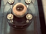

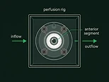
Ex-vivo human donor-eye perfusion; internal, unpublished, not peer reviewed. This is a laboratory model, not a clinical study. It does not establish that OF-01 reduces intraocular pressure in living patients; that must be shown in prospective clinical trials.

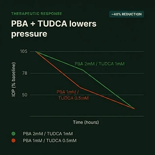
IOP reduction observed in perfused human glaucoma donor eyes with PBA + TUDCA, acting on the conventional outflow pathway rather than by suppressing aqueous production.
Figure is approximate and representative of observed internal data; unpublished and not peer reviewed. Not predictive of human clinical results.
From trabecular stress to hypothesized patient benefit.
The logic chain OF-01 is built on, from disease biology to the benefit it is hypothesized to produce. Everything downstream of the biology is a design goal or a preclinical observation, not an established clinical outcome.
Disease biology
- POAG and steroid-induced ocular hypertension converge on a stressed, fibrotic trabecular meshwork.
- Misfolded-protein load drives chronic UPR and ATF4 signaling.
- Mitochondrial injury, ECM buildup, stiffness and cell loss raise conventional outflow resistance.
Drug rationale
- PBA improves protein folding and suppresses maladaptive UPR.
- TUDCA stabilizes ER and mitochondrial membranes and reduces apoptosis, inflammation and oxidative injury.
- Combination hypothesis: complementary rescue at lower topical exposure.
Molecular design goals
- Lower IRE1, GRP78, protein aggregates and ATF4 / CHOP burden.
- Reduce fibronectin and pathologic ECM deposition.
- Restore TM viability, mitochondrial quality control and physiologic outflow signaling.
Preclinical evidence
- Primary human TM cells: reduced ER stress, aggregation and fibrosis.
- Inducible MYOC glaucoma mouse: IOP reduction with preserved RGCs and PERG.
- Perfused human donor eyes: improved outflow; retinal models report up to ~40% RGC protection.
Hypothesized benefit
- Near-term: durable IOP reduction.
- Intermediate: fewer adjunctive drops, delayed SLT or MIGS escalation.
- Long-term: slower RNFL / GCC thinning and visual-field loss, with possible direct RGC protection.
The combination, donor-eye and mouse results remain preclinical. Prospective clinical trials must establish ocular safety, pharmacokinetics, dose-response, durable IOP reduction, outflow facility, OCT RNFL / GCC and visual-field progression, and treatment burden. No clinical benefit is established.